
Heart & Blood
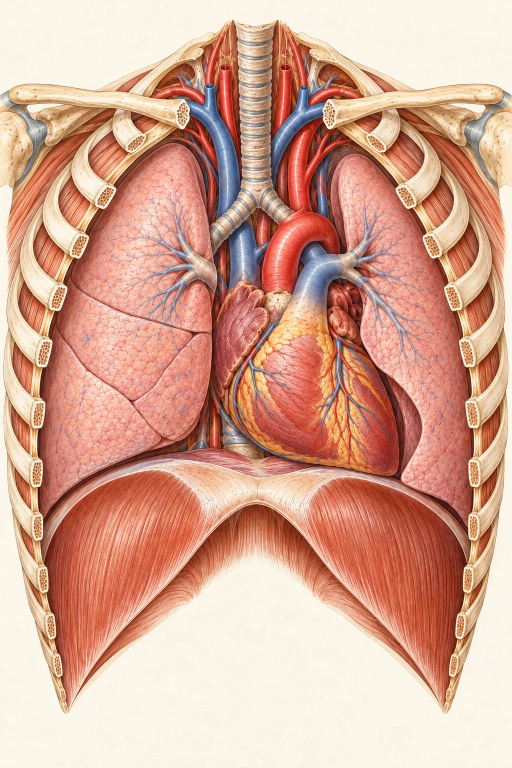
Your heart lies between your lungs. Its pumping action keeps blood moving. Blue on vessels is a diagram convention. Blood is red.
Organ view
Enter this sceneFollow blood through the heart, lungs and body.
Follow circulation through the heart, lungs and body. Explore the chambers, valves and blood vessels, then change scale to discover red blood cells and oxygen exchange. The illustrations simplify anatomy to explain each part of the journey.
Your heart lies between your lungs. Its pumping action keeps blood moving. Blue on vessels is a diagram convention. Blood is red.
Organ view
Enter this sceneThe heart is made mostly of muscle. Vessels on its surface bring blood to the muscle itself. Blue on vessels is a diagram convention. Blood is red.
Organ view
The right side sends blood to the lungs. The left side sends blood to the body. A wall separates them.
The right side sends blood to the lungs. The left side sends blood to the body. A wall separates them. Blue on vessels is a diagram convention. Blood is red.
Teaching cutaway
Large veins bring blood back from the body. This blood enters the right atrium before moving into the right ventricle.
Blood leaves the right ventricle through the pulmonary valve. The pulmonary arteries carry it toward the lungs.
Pulmonary veins bring blood from the lungs into the left atrium. The next passage leads into the left ventricle.
The left ventricle sends blood toward the body. Its muscle wall is thicker than the right ventricle's.
Large veins bring blood back from the body. This blood enters the right atrium before moving into the right ventricle. Blue on vessels is a diagram convention. Blood is red.
Teaching cutaway
Enter this sceneThe tricuspid valve lies between the right atrium and right ventricle. Its leaflets help prevent backward flow. Blue on vessels is a diagram convention. Blood is red.
Teaching cutaway
Enter this sceneBlood leaves the right ventricle through the pulmonary valve. The pulmonary arteries carry it toward the lungs. Blue on vessels is a diagram convention. Blood is red.
Teaching cutaway
Enter this sceneBlood travels through the lungs, then returns to the heart. Airways carry air; blood vessels carry blood. Blue on vessels is a diagram convention. Blood is red.
Organ view
Enter this sceneAlveoli are tiny air sacs. Their thin walls lie beside capillaries where gases can pass between air and blood. Microscopic teaching enlargement. Sizes and wall thickness are simplified.
Microscopic teaching view
Enter this sceneOxygen passes from an air sac into nearby blood. Hemoglobin in red blood cells carries it onward. Carbon dioxide passes the other way. Microscopic teaching enlargement. Sizes and wall thickness are simplified.
Microscopic teaching view
A mature red blood cell has a dip on each side and no nucleus. Hemoglobin inside it carries oxygen.
Pulmonary veins bring blood from the lungs into the left atrium. The next passage leads into the left ventricle. Blue on vessels is a diagram convention. Blood is red.
Teaching cutaway
Enter this sceneThe left ventricle sends blood toward the body. Its muscle wall is thicker than the right ventricle's. Blue on vessels is a diagram convention. Blood is red.
Teaching cutaway
The mitral valve opens during filling. Fine cords connect its leaflets to muscles inside the ventricle.
Blood leaves the left ventricle through the aortic valve. The aorta branches into arteries that carry it onward. An isolated oblique view of the left heart. The camera differs from the whole-heart view.
Oblique left-heart section
Enter this sceneIn tiny capillaries, oxygen passes out toward nearby cells. Red blood cells remain inside the blood vessel. Microscopic teaching enlargement. Sizes and wall thickness are simplified.
Microscopic teaching view
Enter this sceneBlood returns through small veins that join larger veins. It travels back toward the right side of the heart. Microscopic teaching enlargement. Sizes and wall thickness are simplified.
Microscopic teaching view
Enter this sceneOne circuit connects heart and lungs. The other connects heart and body. Together they form a repeating journey. Separate organ vignettes at different scales. Read the route to follow the connections.
Schematic overview
Enter this sceneA mature red blood cell has a dip on each side and no nucleus. Hemoglobin inside it carries oxygen. Microscopic teaching enlargement. Sizes and wall thickness are simplified.
Microscopic teaching view
Enter this sceneRed blood cells can change shape as blood carries them through narrow capillaries. Microscopic teaching enlargement. Sizes and wall thickness are simplified.
Microscopic teaching view
Enter this sceneArteries carry blood away from the heart. Veins carry it toward the heart. Their walls differ to suit their roles. Some veins also have valves. The vein combines cross-sectional and lengthwise views. Colors are schematic.
Teaching section
Enter this sceneThe heart wall includes a thick layer of muscle. A surrounding sac helps protect the heart and lets it move with less friction. Illustrated teaching view. Shapes and colors are simplified.
Teaching section
Enter this sceneDuring ventricular filling, the atrioventricular valves are open. The aortic and pulmonary valves are closed. A simplified phase illustration. These frames do not represent the full heartbeat.
Phase illustration
Enter this sceneAs the ventricles eject blood, the atrioventricular valves are closed. Blood passes through the open outflow valves. A simplified phase illustration. These frames do not represent the full heartbeat.
Phase illustration
Enter this sceneThe mitral valve opens during filling. Fine cords connect its leaflets to muscles inside the ventricle. A simplified phase illustration. These frames do not represent the full heartbeat.
Phase illustration
Enter this sceneDuring ventricular contraction, the mitral leaflets close. Their supporting cords help stop them bending back into the atrium. A simplified phase illustration. These frames do not represent the full heartbeat.
Phase illustration
Enter this sceneElectrical signals coordinate the heartbeat. They begin in the right atrium and pass through the heart in an ordered sequence. Blue on vessels is a diagram convention. Blood is red.
Teaching cutaway
Enter this sceneCoronary arteries supply the heart muscle. Cardiac veins carry blood away from it. Blue on vessels is a diagram convention. Blood is red.
Organ view
Enter this scene